Features
General purpose of a microscope.
Nowadays, there are 4 main options for the appointment:
children's,
educational,
laboratory and
specialized microscopes. At the same time, different options (at least from the first three) may well be combined in one model — for example, the simplest and most inexpensive educational microscopes may well be positioned as children's, and the most advanced as laboratory ones. Here is a detailed description of the different destinations:
— Children's. The most simple and inexpensive microscopes, designed primarily for children who are taking their first steps in the natural sciences (as well as for other undemanding users who do not need particularly advanced functionality). Accordingly, such devices lack advanced features such as focus lock, Keller lighting, video outputs (for digital and opto-digital models), a trinocular with the ability to connect a camera, etc. In addition, the body can be made in bright colours, and in plastic is usually used as the body material. However, many children's microscopes are equipped with turrets for quick re-tuning of magnification, and the total magnification factor may well exceed 600x out of the box and 1000x in the top configuration.
— Educational. Microscopes well suited for teaching applications; sometimes such an appointment is even di
...rectly indicated by the manufacturer. The specific functionality of such models is quite diverse, the type can also be different (both biological and stereoscopic). In general, devices of this specialization occupy an intermediate position between simple and inexpensive children's microscopes and advanced laboratory equipment. At the same time, there are many models that have a combined purpose — "children's / educational" or "training / laboratory". The first variety is simple and inexpensive, for educational purposes it is suitable mainly for school; the second option, in turn, can be useful even at the university faculty of natural sciences.
— Laboratory. The most advanced type of modern microscopes, designed for full-fledged laboratory research and other serious tasks. Accordingly, such models are not cheap, but they provide a high-quality image and, in general, have the most extensive functionality (although the specific set of features, of course, may be different). Among the features found in laboratory microscopes are a movable stage, installation of light filters, 2 types of illumination (lower and upper), Keller illumination, suitability for special microscopy methods (fluorescence, phase contrast), etc.
— Specialized. Microscopes of a specific design and purpose, one way or another different from more traditional models. These differences may vary; accordingly, the specific specialization also differs. So, recently, portable models for smartphones have gained quite significant popularity: with the help of a special clothespin, such a device is attached directly to the gadget opposite the main camera, and the smartphone screen plays the role of an eyepiece. Another popular variety is compact digital microscopes without their own screens, connected to PCs or laptops via USB, and even to smartphones via Wi-Fi (including via the Internet). This also includes professional equipment with a fairly narrow specialization: stereoscopes with special mounts for dental prosthetics, for soldering microcircuits, etc.; microscopes for metallurgical research; devices on a tripod with a remote rod, designed to inspect individual areas on general objects; comparative microscopes for ballistic and trace investigations in forensics; and etc.Lens
—
Zoom lens. Lens with variable magnification. Such optics allow you to smoothly change the overall magnification of the microscope within certain limits, without changing the objective/eyepiece and without even looking up from observations. On the other hand, zoom lenses are more complicated and more expensive than constant magnification optics. Therefore, they are mainly used in stereoscopic microscopes (see "Type"): in the repair, assembly and other tasks for which such devices are used, the ability to smoothly adjust the multiplicity is extremely useful.
— magnification factor. The magnification provided by the lens. This parameter, along with the magnification of the eyepiece, affects the overall magnification of the device (see above). Most biological microscopes (see "Type") are equipped with several different magnification objectives on the turret; this allows you to adjust the degree of magnification as desired by the user. The standard magnification options for such lenses are 4x, 10x, 40x, 100x.
— Achromat. One of the varieties of colour correction used in lenses. The need for colour correction is due to the fact that light of different colours is refracted differently by lenses, and without additional measures, the image in the microscope would be blurred with iridescent stains. Achromatic is one of the simplest types of colour correction; in such optics, colour distortions in yellow and green are corrected
.... Achromatic lenses have simple design and low cost. However the image quality in them is far from perfect: such a lens gives a clear image only in the centre of the image, the width of the sharpness zone is about a third of the total width of the field of view, and red-blue streaks may appear along the edges of the image. However, this is quite enough for general acquaintance, initial training, and often for more serious tasks.
— Planachromat. An improved and improved version of achromatic lenses (see above). Plan achromats provide additional correction of the field curvature, due to which the area of a clearly visible image in such lenses is at least 2/3 of the total width of the field of view, and often even more. It is these lenses that are recommended for serious study and professional use.
— Rim diameter. The size of the thread used to mount the lens. A larger bore usually means a wider lens, which means higher aperture and better image quality. On the other hand, the large size affects the dimensions, weight and cost of optics. In modern microscopes, diameters from 20 to 35 mm are mainly found. Knowing the size of the thread, you can purchase replacement or spare lenses for the device.Eyepiece
—
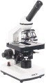
Monocular. An eyepiece with a single lens that can only be viewed with one eye. For obvious reasons, it is only used in biological microscopes (see "Type"). The advantages of monoculars are primarily smaller size and cost than other varieties; in addition, they do not require adjustment for interpupillary distance. On the other hand, constantly looking into the eyepiece with one eye is tiring, so this option is poorly suited for situations where you have to look into the microscope often and for a long time.
—
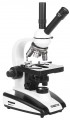
Binocular. Dual eyepiece that can be viewed with both eyes at once. Note that such optics are used not only in stereomicroscopes, originally intended for viewing an object through two lenses (see "Type"), but also in biological microscopes with one lens. The fact is that looking into an optical device with two eyes is much more convenient than with one, while the eyes are less loaded and fatigue does not occur so quickly. Therefore, for serious tasks associated with frequent use of a microscope, binoculars (or trinoculars, see below) are the best option. Such optics cost more than monocular, but this is offset by ease of use.
—
Trinocular. A kind of binocular (see the relevant paragraph), supplemented by a third optical channel for a special camera-video eyepiece. Such a camera is usually connected to a PC or laptop; by installing it in the soc
...ket for the third eyepiece, you can take photos and videos, as well as display the image in real time on the computer screen. At the same time, you can look through the microscope in the usual way. Devices with trinoculars are very functional and versatile, but they are complex and expensive.
— LCD screen. The microscope has an LCD screen that replaces the traditional eyepiece. You do not need to bend over to such a device each time to view the image, which is very convenient if observations need to be combined with record keeping and other similar activities. Microscopes of this design usually have a photo and video function, as well as various built-in tools — for example, a scale grid for estimating the size of visible objects, displayed directly on the screen. In addition, the image on the screen can be seen not only by the direct user, but also by everyone who is nearby; such features are indispensable during training sessions, consultations, presentations, etc. On the other hand, such microscopes turn out to be bulky and expensive.
— magnification factor. The magnification provided by the eyepiece. This parameter, along with the lens magnification, affects the overall magnification of the device (see above). The classic option for eyepieces in microscopes is 10x, but higher values \u200b\u200bare also found. The package may include several eyepieces, of different magnification — to change the overall degree of magnification. There is a multiplicity designation with a letter index, for example, WF10x. This means that the eyepiece has an extended field of view (WF — wide, EWF — extra wide, UWF — extra wide).
— Eyepiece tilt. The tilt of the eyepiece determines the position of the observer's head when looking through the microscope and the overall usability. According to this indicator, three main options can be distinguished: fixed angle, adjustable angle, without tilt. The fixed angle is most often 30° or 45° relative to the horizontal, these values are considered the most convenient. In angle-adjustable microscopes, the entire stand, with tube and stage, is fixed to the base with a swivel mount. This is the most convenient option, allowing you to adjust the tilt to your preference, but the mount tends to become loose over time, so it is rarely used in professional microscopes. The third variety — vertical microscopes, without tilt — have not received much distribution: this design is used in some stereoscopic models (see "Type") in order to ensure that the stage remains strictly horizontal (this is important for some work with microscopic objects).
— Rim diameter. The nominal diameter of the eyepiece used in the microscope, as well as the diameter of the hole in the tube, designed to install the eyepiece. Several standard diameters are used in modern microscopes, in particular 23 and 27 mm. In fact, this parameter is necessary, first of all, if you plan to purchase spare or replacement eyepieces for the microscope, or if you already have an eyepiece on the farm, and you need to evaluate its compatibility with this model.
— Diopter adjustment. The range of diopter correction provided in the eyepiece. This correction is used so that a nearsighted or farsighted person can look through the microscope without glasses or contact lenses. In most models with this function, the correction range is about 5 diopters in both directions; this allows the microscope to be used for low to moderate myopia/farsightedness.Drug agent
Presence of a preparation agent in the design of the object table.
The preparation guide is a device for smooth movement of slides under the microscope lens, as well as fixing the conditional coordinates of individual sections of the preparation. Mechanisms are responsible for the movement, allowing the glass to be shifted separately in the longitudinal and transverse directions. Coordinate fixation is provided by special scales with verniers, the accuracy of determining coordinates can be from 0.1 to 0.01 mm.
This feature is found exclusively in biological microscopes (see "Type"). Its presence can be extremely important for studies involving high magnification factors. Without a slider, the glass would have to be moved by hand, and finding certain areas would be a very difficult, if not impossible, task.
Condenser
Features of the design of the condenser installed in the microscope.
The condenser is part of the illumination system in biological microscopes (see "Type"). This is an optical system that processes the light flux entering the preparation glass in a special way. Different situations may require different ways of doing this; accordingly, different types of condensers can be used in microscopes. However, the most popular nowadays is the simplest Abbe condenser. It ensures the concentration of the beam of light and its uniform distribution over the field of view. Initially, such a device was intended for studies using the bright field method, but it can also be used for phase-contrast observations. The Abbe condenser can be equipped with an iris aperture diaphragm — with its help you can reduce the brightness of the illumination — as well as colour filters.
Other, more specific types of condensers (for example, phase or dark field) are usually purchased separately and are rarely included in the standard microscope equipment.
The characteristics of the condenser may indicate NA — the size of the aperture (active hole) in millimetres, for example, NA \u003d 1.2. This is a rather specific setting; suffice it to say that it is selected by the manufacturer for complete lenses and does not fundamentally affect the choice of a microscope.
Light filters
The presence of
light filters in the scope of delivery of the microscope.
Light filters are installed in the lighting system; they can be interchangeable or built-in (usually on a turret). Anyway, such devices change the characteristics of light, adjusting it to the specifics of the situation. The types and purpose of light filters can be different, as well as their range in the kit; here are some of the most common options:
— Blue colour. Useful in cases where light from an incandescent or "halogen" lamp is used for illumination. Such a filter equalizes the colour temperature (white balance), making the shades of colours colder and providing natural colour reproduction; this is especially important for microphotography, since a properly set white balance is critical to obtaining high-quality images.
— Yellow colour. Kind of the opposite of blue: lowers the colour temperature, giving the image a warmer tint. It is also sometimes useful for adjusting white balance, but yellow filters have another important use: they are well suited for detecting imperfections in metallic surfaces.
— Green colour. Achromatic and planachromatic objectives, which are installed in most modern microscopes, are best at eliminating aberrations in the green part of the spectrum. With this in mind, similar filters are applied: an image painted in a green tint has the least visible distortion. In addition, most objectives
...for phase contrast microscopy are also most effective in the green part of the spectrum (although exceptions are possible).
— Matte (diffuser). White colour filters that do not change the colour of the light, but provide additional dispersion. This can be useful, in particular, when working with low magnification lenses.
— Neutral. Filters in different shades of grey. Used to reduce the intensity of lighting without changing its other characteristics. Such devices can be especially useful when taking photographs — namely, if the camera does not have a sufficiently fast shutter speed. Note that a similar effect can be achieved using a microscope diaphragm, but this is not always the best option when shooting. So, narrowing the aperture reduces the field of view and increases the depth of field (the latter is also not always desirable), while filters do not affect these parameters; besides, in some situations, even the narrowest aperture may not be “dark” enough.
— Light filters for coloured preparations. Improve the visibility of elements painted in a particular colour. Such fixtures are especially popular in biological studies, as they are the most commonly stained specimens and are also the most susceptible to dye fading, making it difficult to view under normal lighting conditions. Note that filters of this type, in contrast to the colour filters described above, do not colour the entire image in a certain colour, but only muffle all other colours, except for their “native”.
— Fluorescent. Filters used in fluorescence microscopy. They are divided into two types — exciting (they separate UV radiation from the general backlight spectrum to illuminate the drug) and closing (protect the user's eyes from ultraviolet radiation and at the same time let the fluorescent glow of the drug pass through).Power source
Methods of nutrition provided in the microscope. Even optical models may require a power source to run the backlight (see above), while for other varieties, power is almost a must. Some models may support multiple power types.
— 230 V network. Connection to a regular 230 V socket. Quite a convenient and practical option, only poorly suitable for portable models (see above).
— USB port. Power supply from the USB connector is often found in digital microscopes (see "How it works"): the device is powered from the same connector through which it is connected to a computer or other external screen. And in optical models, such power supply can be provided in addition to the 230 V network described above. Note that USB ports, among other things, are also found in laptops and other portable devices, which makes it possible to use such microscopes even if there are no outlets nearby. This is especially useful for portable devices (see above).
— Accumulator. Powered by its own built-in battery, in some cases — non-removable. This option makes the microscope completely autonomous and allows you to use it even in the complete absence of external power sources nearby. On the other hand, this moment is relevant mainly for portable models, and then only in some cases, and the built-in battery noticeably affects the weight, dimensions and price of the device. Therefore, purely cordless microscopes are extremely rare, more often this method of power supply is pro...vided in addition to the 230 V network or USB (see above) — as a spare in case of problems with external power.
— Batteries. Another type of autonomous power supply, along with the batteries described above. The presence of a battery compartment is cheaper than the built-in battery, but the batteries themselves have to be purchased separately — and either regularly buy disposable cells, or pay a rather large amount for batteries and a charger for them. In addition, the quality of batteries is highly dependent on the specific brand, and not all cells can normally “start” the microscope and provide an acceptable battery life. Therefore, such power, like battery power, is rare in its pure form, more often it complements the connection to a 230 V network or USB.