Features
General purpose of a microscope.
Nowadays, there are 4 main options for the appointment:
children's,
educational,
laboratory and
specialized microscopes. At the same time, different options (at least from the first three) may well be combined in one model — for example, the simplest and most inexpensive educational microscopes may well be positioned as children's, and the most advanced as laboratory ones. Here is a detailed description of the different destinations:
— Children's. The most simple and inexpensive microscopes, designed primarily for children who are taking their first steps in the natural sciences (as well as for other undemanding users who do not need particularly advanced functionality). Accordingly, such devices lack advanced features such as focus lock, Keller lighting, video outputs (for digital and opto-digital models), a trinocular with the ability to connect a camera, etc. In addition, the body can be made in bright colours, and in plastic is usually used as the body material. However, many children's microscopes are equipped with turrets for quick re-tuning of magnification, and the total magnification factor may well exceed 600x out of the box and 1000x in the top configuration.
— Educational. Microscopes well suited for teaching applications; sometimes such an appointment is even di
...rectly indicated by the manufacturer. The specific functionality of such models is quite diverse, the type can also be different (both biological and stereoscopic). In general, devices of this specialization occupy an intermediate position between simple and inexpensive children's microscopes and advanced laboratory equipment. At the same time, there are many models that have a combined purpose — "children's / educational" or "training / laboratory". The first variety is simple and inexpensive, for educational purposes it is suitable mainly for school; the second option, in turn, can be useful even at the university faculty of natural sciences.
— Laboratory. The most advanced type of modern microscopes, designed for full-fledged laboratory research and other serious tasks. Accordingly, such models are not cheap, but they provide a high-quality image and, in general, have the most extensive functionality (although the specific set of features, of course, may be different). Among the features found in laboratory microscopes are a movable stage, installation of light filters, 2 types of illumination (lower and upper), Keller illumination, suitability for special microscopy methods (fluorescence, phase contrast), etc.
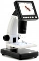
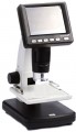
— Specialized. Microscopes of a specific design and purpose, one way or another different from more traditional models. These differences may vary; accordingly, the specific specialization also differs. So, recently, portable models for smartphones have gained quite significant popularity: with the help of a special clothespin, such a device is attached directly to the gadget opposite the main camera, and the smartphone screen plays the role of an eyepiece. Another popular variety is compact digital microscopes without their own screens, connected to PCs or laptops via USB, and even to smartphones via Wi-Fi (including via the Internet). This also includes professional equipment with a fairly narrow specialization: stereoscopes with special mounts for dental prosthetics, for soldering microcircuits, etc.; microscopes for metallurgical research; devices on a tripod with a remote rod, designed to inspect individual areas on general objects; comparative microscopes for ballistic and trace investigations in forensics; and etc.Magnification
The range of magnifications provided by the device is from minimum to maximum.
The magnification of the microscope is calculated by the formula "the magnification of the eyepiece is multiplied by the magnification of the objective." For example, a 20x objective with a
10x eyepiece will give a magnification of 10*20 = 200x. Modern microscopes can be equipped with multi-objective turrets, zoom lenses (see below) and interchangeable eyepieces — so that in most models the magnification can be adjusted. This allows you to adjust the device to different situations: when you need to consider small details, a high degree of magnification is used, but to expand the field of view, the magnification must be reduced.
Detailed recommendations on optimal multiplicities for different tasks can be found in special sources. Here we note that many manufacturers go to the trick and indicate the maximum value of the magnification by the degree of magnification achieved with an additional Barlow lens. Such a lens can indeed give a serious increase in magnification, but it is not a fact that the image will turn out to be of high quality; for more details, see "Complete set".
Research method
Research methods applicable to this microscope model.
— Bright field. The most famous and widely used method of light microscopy. The object under consideration in such studies is placed on a light background, against which it looks darker. Note that different methods of illumination can be used for research: direct through, oblique, reflected. The first option (when the light from a lamp or a mirror under the stage shines through the sample) is optimal for studying transparent samples, the key details of which are darker than the general background. Typical examples are thin sections of animal and plant tissues. Oblique light is similar in application specifics, while it gives a grey background and is inferior to direct light in terms of backlight efficiency, but provides a more embossed image. As for reflected light, in this case it is indispensable when examining opaque objects: samples of ores and other materials, semiconductor wafers, etc. Anyway, bright-field microscopy well reveals, first of all, details that are noticeably different in light transmission or refractive index from the surrounding background (with through illumination), or give noticeable reflections / shadows (with reflected light).
— Dark field. A kind of opposite of bright-field research: the object under consideration or its individual elements are lighter than the surrounding background. However, this is not just a “negative” of the image, but a separate method with its own c...haracteristics. Illumination in dark-field microscopy is usually through, but it is carried out in a specific way: the middle of the light beam is blocked by a hood, and the light “cylinder”, passing through the condenser lens, turns into an “hourglass”. At the same time, in the narrowest place of such a "clock" there is a preparation, and towards the lens, the light cone expands so that it does not fall into the optics. Thus, the user sees in the microscope only the light scattered by the preparation and the dark background around. This method of research, among other things, allows you to identify "smooth" details that do not stand out sharply against the surrounding background and are not visible in a bright-field study. Among the applications of dark-field microscopy are the work with unstained biological preparations (cells, tissue samples, microorganisms), as well as the study of some transparent materials for small surface defects.
— Phase contrast. A method used to study transparent and colourless objects with an inhomogeneous structure, used when this inhomogeneity cannot be detected by more traditional bright-field microscopy. The idea of this method is that when passing through structures with different refractive indices, light receives different phase changes. These changes are not visible with ordinary optics, but they can be made visible with the help of special equipment — namely, a condenser and a lens of a special design. Accordingly, such equipment is necessarily included in the scope of delivery of the microscope.
— Fluorescent. This method provides the illumination of observed objects with light of a certain wavelength, under the influence of which these objects or their individual elements begin to glow, while the background remains dark. If necessary, coloring substances are introduced into the preparation to improve luminosity (a typical example is biological objects, most of which fluoresce rather weakly by themselves). For illumination, usually, UV radiation is used, therefore this method is also called ultraviolet microscopy; The image enters the eyepiece of the microscope through a filter that filters out UV rays, but freely passes the visible glow of the drug.
One of the main features of fluorescence microscopy is high resolution: it allows you to clearly see even very small objects that are not visible in the usual visible range. In fact, this method in terms of resolution is between classical optical and electron microscopy; At the same time, in contrast to electron and atomic microscopes, devices with the support of the UV method make it possible to examine even the hardware of living cells and microorganisms. And some special variants of this technique make it possible to achieve not micro, but nanoscopic magnifications. The second popular use of fluorescence microscopy is the detection of particles, elements, inclusions, etc., which are not visible under ordinary light, but stand out well in ultraviolet light. A typical example is the surface of many metals and alloys.
Object table
The design of the object stage provided in the microscope.
— Stationary. Subject table, fixed motionless; focus in such microscopes is carried out by moving up and down the tube with the objective and the eyepiece. Such systems are simple and inexpensive, but focus while looking through a constantly moving eyepiece is not very convenient. In addition, for advanced biological microscopes (see "Type") with binoculars and trinoculars (see "Eyepiece"), this option is also poorly suited for some design reasons. But the vast majority of stereomicroscopes are equipped with stationary tables — this is the most reasonable design, taking into account the specifics of the application.
—
Movable. In microscopes of this type, the entire optical system is fixedly fixed on a tripod, and the stage can be moved up and down to focus the optics. This design is found exclusively in biological microscopes (see "Type"). It is somewhat more complicated and expensive than with a fixed table, but at the same time it is much more convenient: when focus, the eyepiece does not move, which allows you to comfortably adjust the image without looking up. In addition, it is the movable stage that is most suitable for advanced devices with binoculars and trinoculars (see "Eyepiece"), almost all such microscopes have such equipment.
Focus
Types of focus (focus) provided in the microscope. Focus is carried out by changing the distance between the object under consideration and the lens; its types can be:
— Rough. This method means that there is one rotary control responsible for moving the lens or stage. The advantages of this design are simplicity and low cost. At the same time, focus at high magnifications in such microscopes is a rather difficult task: you have to turn the tuning knob literally in fractions of a millimetre.
—
Coarse / Fine. Focus, carried out by two mechanical controls — for preliminary focus and for final fine tuning. Such a tuning is more convenient in itself than only a rough one (see above), and at high magnifications it can be simply irreplaceable. On the other hand, the presence of an additional regulator complicates and increases the cost of the design, so this option is found mainly in semi-professional and professional microscopes.
— Manual. A method that assumes the absence of a focus mechanism as such. Focus in such devices is carried out due to the fact that the user manually moves the lens — for example, moving it up and down on a vertical tripod and fixing it in the desired position with a clamp, or tilting it back and forth on a swivel mount. This option is only suitable for models with a low magnification that do not require special accuracy when focus; it is found mainly in digital microscopes without thei
...r own screen (see "How it works"), as well as portable models (see the relevant paragraph).